EMB
Every tooth has two anatomical parts: the crown and the root. The crown is the part of the tooth that is normally visible in the mouth (above the gumline), while the root of a tooth is the unseen part embedded in the jawbone (below the gumline). The gumline is a smooth surface cavity that separates the gum from the exposed parts of the tooth.
Usually, dentists extract the tooth along with its root(s), yet sometimes the root tips may fracture during the extraction process. Additionally, when the tooth is damaged through accident or decay, the root fragments may be retained within the jawbone and gums, causing problems such as mouth infections and pain. On the one hand, the prevalence of retained root fragments is reported to be 11-37%; on the other hand, between 10-20% of extractions become “surgical” even though initially it has seemed possible to remove the retained root fragments without encountering any complications.
Root extraction can prove problematic for dental practitioners as surgery also heightens the chance of a number of complications. Therefore, we are proposing a dental instrument that helps remove retained roots, preventing the need to do surgery and consequently the common postoperative complications.
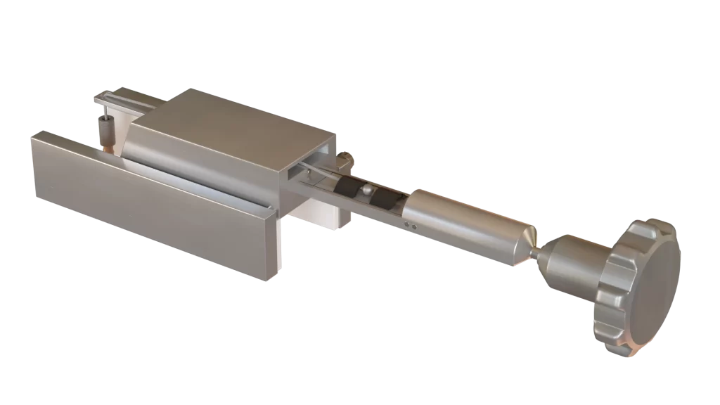
We recommend a practical and effective dental instrument to remove broken teeth roots and prevent operative and postoperative complications. Expandable Micro Bur (EMB) is a functional dental instrument used to remove broken teeth roots that cannot be extracted by routine methods. The device consists of a spacers, needles, bur base, a round bur that drills halfway through the root canal, expands to make a spherical cavity around itself, and acts as an efficient extraction-aiding anchor. The utilization of EMB would introduce a new technique in removing broken teeth roots by which surgical trauma and post-extraction disorders are minimized. Using EMB would also minimize the amount of invasion to the surrounding tissues, removing the need for intense pressure that the practitioner usually applies.

How it works

This procedure is done in 5 steps:
- Pre-operative: This step depicts a broken root stuck in its socket before the operation.
- Drilling: Using a micro-motor, a round bur drills approximately halfway through a root canal, similar to a conventional bur while spacers are on.
- Screwing: spinning stops, the micro-motor is detached from EMB; round bur is unscrewed from the bur base; spacers are loosened or removed (depending on the expansion degree required); round bur is placed back into the canal; bur base is mounted on micro-motor, with the help of a needle holder; round bur and bur base are screwed back. Round bur carves its periphery area, making a spherical cavity around itself. This step would take only a short while.
- Expansion: after the spinning stops, the micro-motor is detached while EMB remains in place. As the split round bur expands in the root canal, it acts as an efficient extraction-aiding anchor.
- Anchoring: The firm anchor eases the extraction of the broken root with the help of a needle holder.
- Extraction: the dentist extracts the root without complications.

EMB PRO
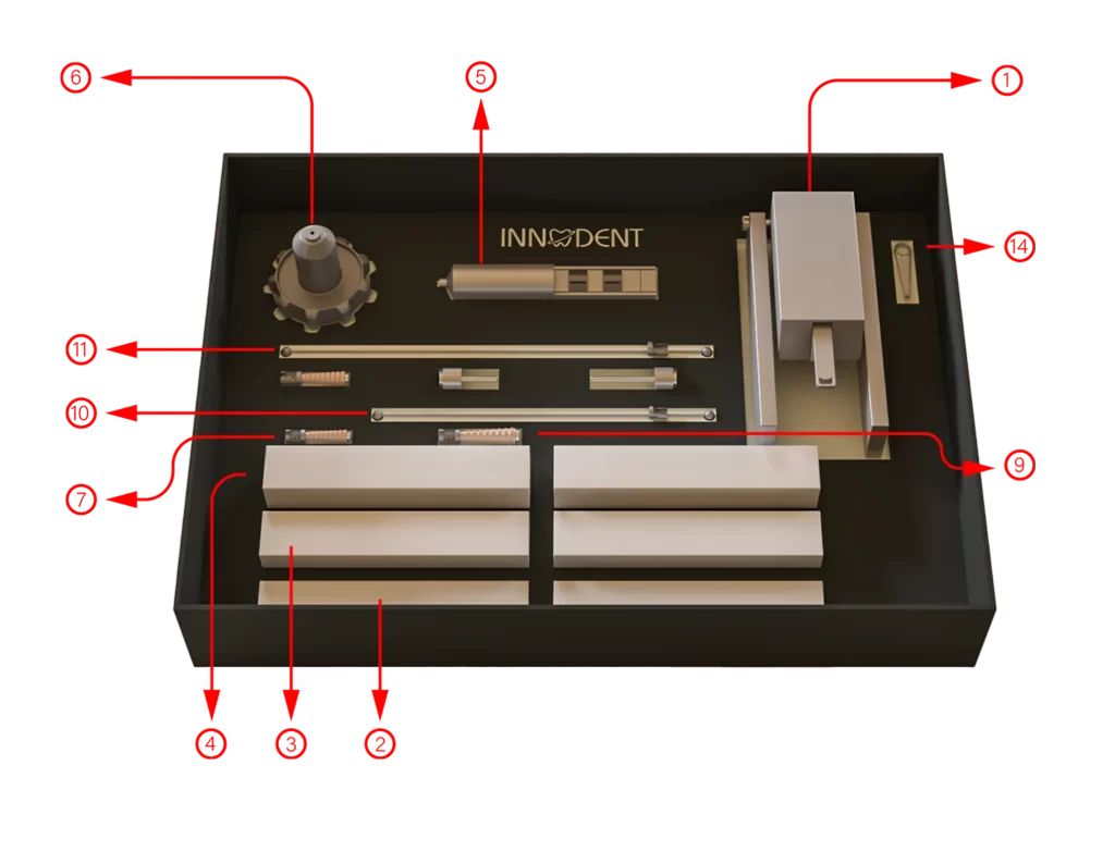
We are excited to announce that we have released an enhanced version of our EMB product called EMB Pro. With the new design, there is no longer a need to drill the retained root with the device, as the bur has been designed to fit into pre-drilled holes, and its head can expand to lock into the location and form a firm grip on the dental root. The new mechanism has eliminated the need to use separate bur pieces. The rest of the instrument is solely designed to ease the process of extracting a retained tooth root. Also, the device reduces the force required for the operation. This reduction in force would also prevent the dealing of further damage to the tissue surrounding the root being extracted and the adjacent teeth. Our new device can still be used on all teeth as it is retractable and has adjustable placement support bars that fill in the gap between the device and the teeth it is being placed on. EMB Pro is still centered around the painless extraction of retained root fragments. The product’s new design allows the user to adjust the amount of expansion required to form a firm grip on the retained root fragment, which enables the formation of an optimizable anchor. As some of the retained root fragments might have been subject to decay, the amount of direct pressure they can withstand can vary depending on their state. An adjustable expansion would allow the practitioner to control the pressure conveyed through the bur directly.
How it works
A hole must be drilled inside the retained root fragment using the already available standard dental drills in the dental clinic. EMB Pro’s dental bur is placed inside the drilled hole. Using the provided screw that can be locked on top of the bur’s head, EMB Pro would expand to fit perfectly inside the hole, forming an anchor. The screw is then detached from the bur. Before attaching the extraction device’s main body to the rails, the appropriate placement support bar based on the size of the tooth being treated must be placed on the rails. The rails have an indentation that the support bars can be fit into. An extraction device is placed on top of the retained root fragment and the other two adjacent teeth. The device is locked onto the bur’s head. It must then be adjusted in accordance with the diameter of the teeth it has been placed on using the provided screw that is placed on the side of the device. Furthermore, the plate on top of the extraction device must also be adjusted to form a 90-degree angle using the 4mm screws on the back of the main device. The rope placed where the handle is attached to the main part of the device has a spherical structure that allows it to become latched in either one of the bridges on the handle, depending on the required height in relation to the EMB Pro. The large screw placed at the back of the handle must be twisted to extract the root fragment.
RCD
Problem
A root canal treatment is a common dental procedure that treats the pulp of a tooth. The success of this operation depends on several factors, including, but not limited to, identifying and assessing the DL (Disto-Lingual) curvature of a root. Radiographic examinations are often used for this purpose; however, this method presents some major limitations:
- Creating a ‘shadowgraph’ produces only two-dimensional images in the process of compressing three-dimensional anatomical images.
- It is necessary to have a mesial and/or distal parallax view to get additional information that otherwise would be inaccessible due to the verticality of the radiographic images.
- Heightening the risk of an inaccurate diagnosis of the DL curvature of the roots because of their unique anatomical shape and X-ray imagining limitations.
In the past decade, several nickel-titanium instruments, as well as rotary endodontic handpieces have been developed for manual root canal preparation. These instruments were designed to improve the overall treatment results and to simplify the difficult and time-consuming process of cleaning, shaping, and enlarging the root canal system. We offer a solution that can help identify the root curvature precisely and minimize anatomical challenges and limitations of current endodontic approaches.
Solution
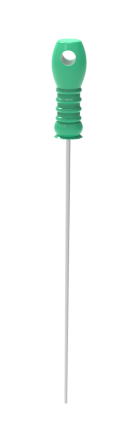
Our product, “Root Curve Detector” or RCD, is a piece of a dental instrument designed to solve the issue of accurately determining the curvature and direction of the root canal. Our cost-effective solution is used together with the radiographic images to increase the precision of endodontic operation.
RCD is a calibrated product made from Nickel-Titanium that has a shape similar to endo files or spreaders. It has cross-sectional grooves that are created at equal intervals of 0.1 mm in a range of 6 mm starting from the beginning of its tip. Using RCD, the curvature of a root canal can be determined when taking X-Ray images by placing this tool inside the canal. A buccal or lingual curvature can be detected based on the changes in the RCD’s degrees.
In the case of a buccal curve, the distance between the scalings on the RCD will be increased; whereas a lingual curve will lead to a decreased distance.
RCD helps dentists determine whether a curvature is forward or backward which is something that was not previously visible in X-ray images due to the verticality of the radiographic images. It would also allow documenting and confirming the accuracy of the dentist’s treatment, which is a vital procedure in regulations and policies in several countries. It can be confidently said that RCD provides a cost-effective method that facilitates determining severe root curvatures before performing a root canal treatment.